
Breakthroughs in the Field of Human Ear Research
From the creation of the first Otopathology Laboratory in the United States in 1924, the esteemed Network of Human Temporal Bone Laboratories has been at the forefront of groundbreaking discoveries in human ear research. This work has deepened our understanding of hearing and balance disorders, transforming the way these conditions are diagnosed and treated. Below, we showcase just a few examples of past and present studies that demonstrate the remarkable impact and potential of temporal bone research.
Stay Up-To-Date With the Latest Advancements in Human Ear Research
Johns Hopkins University
Creation of First Otopathology Laboratory in the U.S.
In 1924, Dr. Samuel Crowe, MD, first chair of the Department of Otolaryngology at Johns Hopkins Hospital, created the first otopathology laboratory in the United States. He appointed Dr. Stacy Guild, an anatomist from University of Michigan, as the laboratory director. Together, they collected more than 1,800 temporal bones between 1924 and 1938, enabling the development of histological techniques for decalcification, embedding, staining, and analysis of cochlear structures—methods still in use today. Their pioneering work made possible the correlation of histological findings with audiometric tests—a groundbreaking technology at the time—and vestibular function in patients. This advancement led to major discoveries such as the first explanations of cochlear tonotopy, the cellular basis of presbycusis, and one of the earliest classifications of hearing loss severity.
Johns Hopkins University
Max Brödel’s 1939 Classic Coronal Illustration of the Inner Ear
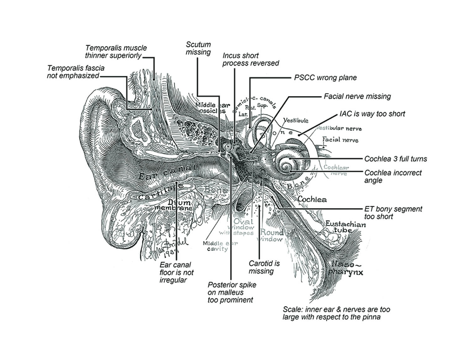
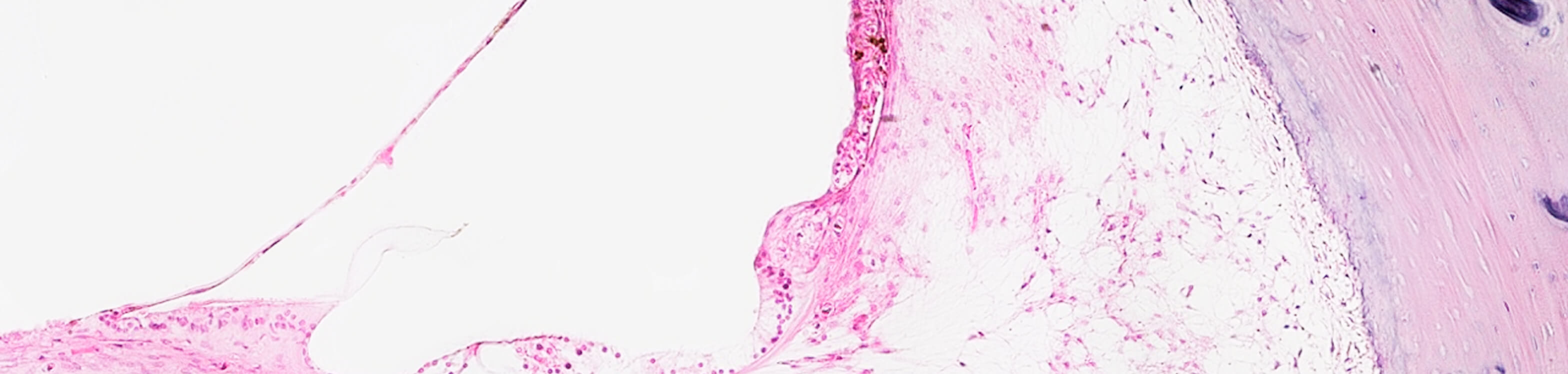
In 1941, Max Brödel, Director of the Department of Art as Applied to Medicine at Johns Hopkins School of Medicine, completed what remains the most famous illustration of the inner ear: a vertical plane representation of the temporal bone, parallel to the superior semicircular canal, created using the laboratory’s extensive collection of histologically sectioned specimens (Illustration published posthumously in 1946).
Notable Publications:
UCLA
Immunohistochemical Protocols
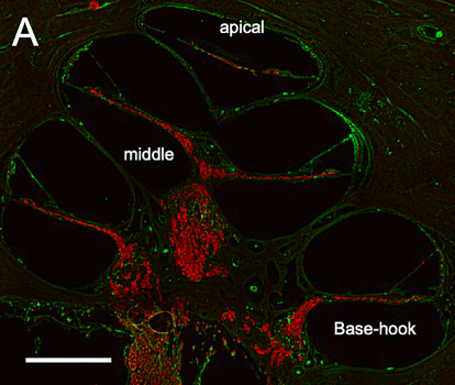
The NIDCD National Temporal Bone Laboratory at UCLA standardized immunohistochemical protocols and methods to study the normal and pathological human inner ear. Given the scarcity of tissue available and the high cost of processing the human temporal bones using the traditional methods we proposed alternative choices such as microdissection, frozen and paraffin sectioning. To optimize the use of invaluable celloidin embedded sections of the human temporal bone our laboratory and others, have been developing methods to remove celloidin from the tissue sections and subsequent antigen retrieval. The best process and preserved tissue methods are being shared with inner ear basic researchers to corroborate their previous findings in animal models. We describe immunofluorescence protocols for allow the identification of several antigens in the same sections.
Notable Publications:
UCLA
National Temporal Bone Laboratory
Prior to the formation of the NIDCD National Human Ear Resource Network in 2023, the NIDCD National Temporal Bone Laboratory at UCLA spearheaded the creation of a national and international network of inner ear researchers that study human temporal bone sections and microdissected organs. A number of groundbreaking publications resulted from this collaboration, including:
- Characterizing Adult Cochlear Supporting Cell Transcriptional Diversity Using Single-Cell RNA-Seq: Validation in the Adult Mouse and Translational Implications for the Adult Human Cochlea
- Mouse Models of Human Pathogenic Variants of TBC1D24 Associated with Non-Syndromic Deafness DFNB86 and DFNA65 and Syndromes Involving Deafness
- Identification of a genetic variant underlying familial cases of recurrent benign paroxysmal positional vertigo
- Emerging Mechanisms in the Pathogenesis of Menière’s Disease: Evidence for the Involvement of Ion Homeostatic or Blood–Labyrinthine Barrier Dysfunction in Human Temporal Bones
- Single-cell transcriptomic atlas reveals increased regeneration in diseased human inner ear balance organs
University of Minnesota
Paparella Otopathology & Ear Pathogenesis Laboratory: Otitis Media
Founded in 1963 by Dr. Michael Paparella, the University of Minnesota’s Paparella Otopathology & Ear Pathogenesis Laboratory has made groundbreaking contributions to the field of otology and neurotology, pioneering research that advances our knowledge of hearing and balance disorders. Notably, the Lab spearheaded research that demonstrated the hearing and vestibular sequelae secondary to otitis media, and introduced the theory of the otitis media continuum, which has reshaped our understanding of pathophysiological mechanisms of otitis media. The lab also introduced the concept of silent otitis media, highlighting how the condition can progress without noticeable symptoms.
Notable Publications: